DIAGNOSIS

Diagnosing glaucoma isn’t just about checking eye pressure. While high intraocular pressure is a key factor, it alone doesn’t confirm the disease some people with high pressure never develop glaucoma, and many with glaucoma have normal pressure.

To make an accurate diagnosis, your ophthalmologist will look closely at your optic nerve, measure your eye pressure, and assess your vision to see if any damage has occurred 

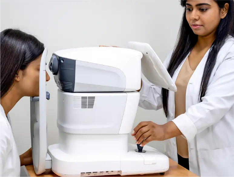
An eye care specialist will review your medical history and perform a comprehensive eye examination.

This may include a series of diagnostic tests, such as:

Tonometry (Measuring Intraocular Pressure)

This test checks the internal pressure of your eye, known as intraocular pressure (IOP), which can indicate the risk of glaucoma.

TONOMETRY1,2

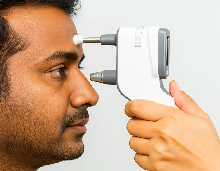
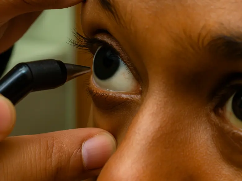
Tonometry is a key test used to measure intraocular pressure (IOP), which, when elevated above 21 mmHg, is a major risk factor for glaucoma a progressive eye disease that can lead to permanent vision loss. The goal is to monitor and manage IOP to prevent or slow glaucoma progression through timely treatment such as eye drops, laser therapy, or surgery.

During the test, the eye is numbed with drops, sometimes with a dye for better visibility, and the pressure is measured using a gentle touch or a puff of air.

References: 1. Bader J, Zeppieri M, Havens SJ. Tonometry. 2023 Dec 12. In: StatPearls [Internet]. Treasure Island (FL): StatPearls Publishing; 2025 Jan–. PMID: 29630277.
  1. Stevens S, Gilbert C, Astbury N. How to measure intraocular pressure: applanation tonometry. Community Eye Health. 2007 Dec;20(64):74-5. Erratum in: Community Eye Health. 2008 Jun;21(66):34. PMID: 18330450; PMCID: PMC2206330.

OPHTHALMOSCOPY (OPTIC NERVE ASSESSMENT)

Also known as a dilated eye exam, this involves inspecting the optic nerve’s shape and color at the back of the eye for any signs of damage.

OPTHALMOSCOPY (OPTIC NERVE ASSESSMENT)1

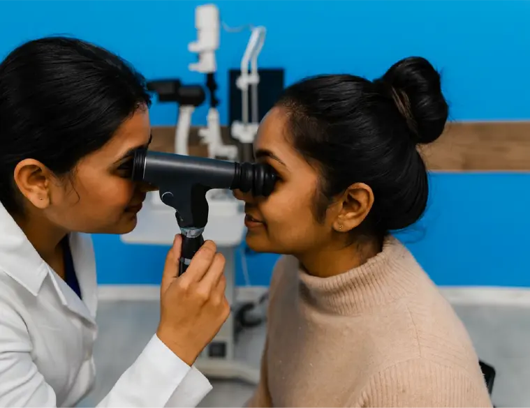
Ophthalmoscopy, or fundoscopy, is a test that allows doctors to examine the inner back part of the eye (the fundus) to detect various eye conditions. Using an ophthalmoscope, which shines light and magnifies internal structures, the doctor can view the retina, optic nerve, choroid, and blood vessels.

Dilating drops are used to widen the pupil for a clearer view. The doctor then checks the optic nerve for signs of glaucoma related damage and examines the retina and blood vessels for any abnormalities. Though the bright light may cause slight discomfort, the procedure is safe, quick, and painless and usually takes 5 to 10 minutes. 

References: 1. Steele A. Diagnosing Glaucoma. The Glaucoma Foundation [Internet]. New York: The Glaucoma Foundation;[cited 2025 Sep 2]. Available from: https://glaucomafoundation.org/about-glaucoma-2/diagnosing-glaucoma/
  1. MedlinePlus Medical Encyclopedia [Internet]. [cited 2025 Sep 2]. Available from: https://medlineplus.gov/ency/article/003881.htm#:~:
text=Direct%20ophthalmoscopy
  1. Fundoscopy (Ophthalmoscopy)[Internet]. Cleveland Ohio: Cleveland Clinic; [cited 2025 Sep 2]. Available from: https://my.clevelandclinic.org/health/diagnostics/ fundoscopy-ophthalmoscopy
  2. Testing for Glaucoma Australia [Internet].[cited 2025 Sep 2]. Available from: https://glaucoma.org.au/what-is-glaucoma/testing-for-glaucoma

Perimetry (Visual Field Analysis)

This test maps your entire field of vision to detect any blind spots or peripheral vision loss, which are common signs of glaucoma.

PERIMETRY (VISUAL FIELD ANALYSIS)1,2,3,4

Loss of peripheral vision is a common symptom of glaucoma and can be detected using a Perimetry test.

Perimetry, or a visual field test, assesses how well you can see across your entire field of vision both central and side vision. During the test, you’ll focus on a central point on a screen while small lights flash in various areas. Each time you see a light, you press a button, helping the doctor map how much of your vision is working properly.

References: 1. Anderson DR. Standard perimetry. Ophthalmol Clin North Am. 2003 Jun;16(2):205–12, vi. doi: 10.1016/s0896-1549(03)00005-1. PMID: 12809158.
  1. American Academy of Visual field testing [Internet]. San Francisco: AAO; [cited 2025 Sep 1]. Available from: https://www.aao.org/eye-health/tips-prevention/visual-field-testing
  2. Glaucoma Research Testing for glaucoma [Internet]. San Francisco: Glaucoma Research Foundation; [cited 2025 Sep 1]. Available from: https://glaucoma.org/understanding-glaucoma/testing
  3. The Glaucoma Diagnosing glaucoma [Internet]. New York: The Glaucoma Foundation; [cited 2025 Sep 1]. Available from: https://glaucomafoundation.org/about-glaucoma-2/diagnosing-glaucoma/

GONIOSCOPY (DRAINAGE ANGLE EVALUATION)

This procedure examines the angle where the iris and cornea meet, helping the doctor determine whether the eye’s drainage system is functioning properly.

GONIOSCOPY (DRAINAGE ANGLE EVALUATION)1

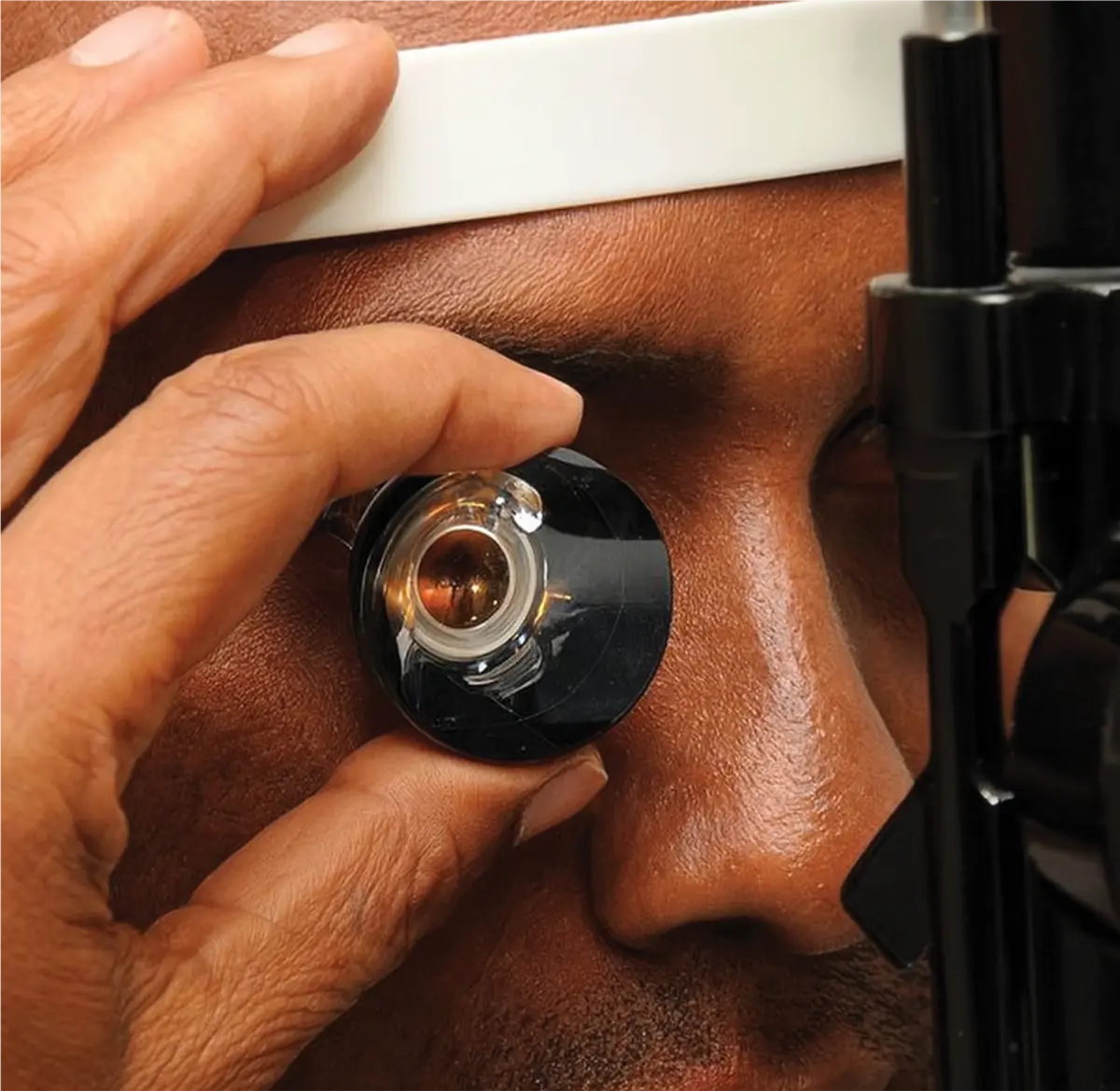
Gonioscopy is a test used to examine the eye’s drainage angle, where fluid exits the eye. Since the eye continually produces fluid, it’s essential that it drains properly through this angle. This test helps determine if high eye pressure is due to a blocked or closed angle (angle-closure glaucoma) or an open but poorly functioning angle (open-angle glaucoma), which is crucial for deciding treatment.

During the procedure, numbing drops are applied, and a special mirrored lens is gently placed on the eye for examination

References: 1. Nolan W, Onakoya A. Gonioscopy skills and techniques. Community Eye Health. 2021;34(112):40-42. Epub 2022 Jan 31. PMID: 35210702; PMCID: PMC8862628.

PACHYMETRY (CORNEAL THICKNESS MEASUREMENT)

This test measures the thickness of your cornea, which can influence eye pressure readings and affect glaucoma risk assessment.

PACHYMETRY (CORNEAL THICKNESS MEASUREMENT) 1,2,3

Corneal thickness is an important risk factor for glaucoma, as thinner cornea can result in higher intraocular pressure readings, increasing the risk of the disease. To assess this, a simple and painless test called Pachymetry is used.

Pachymetry is typically done with an ultrasound device or laser scanner that uses sound waves or light to measure corneal thickness at specific points. Knowing your corneal thickness helps your eye doctor make more accurate decisions for early detection and better management of glaucoma.

References: 1. Anderson DR. Standard perimetry. Ophthalmol Clin North Am. 2003 Jun;16(2):205–12, vi. doi: 10.1016/s0896-1549(03)00005-1. PMID: 12809158.
  1. American Academy of Visual field testing [Internet]. San Francisco: AAO; [cited 2025 Sep 1]. Available from: https://www.aao.org
/eye-health/tips-prevention/visual-field-testing
  1. Glaucoma Research Testing for glaucoma [Internet]. San Francisco: Glaucoma Research Foundation; [cited 2025 Sep 1]. Available from: https://glaucoma.org/understanding-glaucoma/testing
  2. The Glaucoma Diagnosing glaucoma [Internet]. New York: The Glaucoma Foundation; [cited 2025 Sep 1]. Available from: https://glaucomafoundation.org/about-glaucoma-2/diagnosing-glaucoma/